Fluorescence: Difference between revisions
Jump to navigation
Jump to search
No edit summary |
|||
| (9 intermediate revisions by the same user not shown) | |||
| Line 5: | Line 5: | ||
*[[Imaging optics module (IOM)]] | *[[Imaging optics module (IOM)]] | ||
*[[Sample module (SM)]] | *[[Sample module (SM)]] | ||
*[[NPI Manuals]] | |||
*[[media:nanoPi_usersmanual_2007_07_20.pdf|Manual for Nanoprobe Instrument]] | *[[media:nanoPi_usersmanual_2007_07_20.pdf|Manual for Nanoprobe Instrument]] | ||
===Detectors=== | ===Detectors=== | ||
*[[Vortex ME4 (SII 4-element silicon drift diode)]] | *[[Vortex ME4 (SII 4-element silicon drift diode)]] | ||
*[[Vortex (SII 1-element silicon drift diode)]] | *[[Vortex (SII 1-element silicon drift diode)]] | ||
*[[Ge detector (Canberra single element LEGe)]] | *[[Ge detector (Canberra single element LEGe)]] | ||
*[[Region of Interest (ROI)]] | |||
===Optics=== | ===Optics=== | ||
*[[Focusing zone plates]] | *[[Focusing zone plates]] | ||
| Line 15: | Line 19: | ||
*[[Fluorescence Standard Reference Material]] | *[[Fluorescence Standard Reference Material]] | ||
===Analysis=== | ===Analysis=== | ||
Instruction to [[media:Import_Fluo_scans_into_Origin.pdf|import 2D fluorescence images into Origin]]. | Instruction to [[media:Import_Fluo_scans_into_Origin-1.pdf|import 2D fluorescence images into Origin]]. | ||
===Coordinate Systems=== | ===Coordinate Systems=== | ||
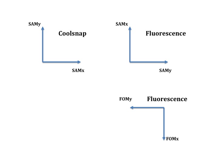
The relation between the coordinate system of the [[Coolsnap]] camera and the y-x-scans in scanView is shown in this [[media:Fluorescence vs transmission orientation.jpg|picture]]. | The relation between the coordinate system of the [[Coolsnap]] camera and the y-x-scans in scanView is shown in this [[media:Fluorescence vs transmission orientation.jpg|picture]]. <br/> | ||
Take Coolsnap transmission image and rotate it by 270 degree. Now flip horizontal. This gives you the orientation of HPiezoY-X scans. | |||
===Links=== | |||
[http://xdb.lbl.gov X-ray Data Booklet] | |||
[[image:DataBook_Image_Small.jpg]]<br/> | |||
[http://henke.lbl.gov/optical_constants/ X-ray interactions with matter]<br/> | |||
[[Category:XMG]][[Category:Fluorescence]][[Category:Controls]] | [[Category:XMG]][[Category:Fluorescence]][[Category:Controls]] | ||
Latest revision as of 22:56, November 8, 2009
Back to X-Ray Microscopy
Nanoprobe Instrument
- Focusing optics module (FOM)
- Condenser module (CM)
- Imaging optics module (IOM)
- Sample module (SM)
- NPI Manuals
- Manual for Nanoprobe Instrument
Detectors
- Vortex ME4 (SII 4-element silicon drift diode)
- Vortex (SII 1-element silicon drift diode)
- Ge detector (Canberra single element LEGe)
- Region of Interest (ROI)
Optics
Standards
Analysis
Instruction to import 2D fluorescence images into Origin.
Coordinate Systems
The relation between the coordinate system of the Coolsnap camera and the y-x-scans in scanView is shown in this picture.
Take Coolsnap transmission image and rotate it by 270 degree. Now flip horizontal. This gives you the orientation of HPiezoY-X scans.
